

Uterine fibroids are a common health concern, and medical advancements now offer less invasive alternatives that prioritize patient recovery and organ preservation. Interventional radiology provides effective solutions for managing fibroid symptoms without the need for major surgery. This approach utilizes advanced imaging guidance to perform minimally invasive procedures, offering a viable option for those seeking relief. Understanding the nature of fibroids and the specific techniques available helps patients make informed decisions about their healthcare.
What Are Uterine Fibroids?
Uterine fibroids are noncancerous growths of the uterus that often appear during childbearing years. These growths are also called leiomyomas or myomas, and they almost never develop into cancer. They range in size from undetectable seedlings to bulky masses that distort and enlarge the uterus.
Interventional radiology plays a pivotal role in treatment strategies. This medical specialty focuses on using minimally invasive image-guided procedures to diagnose and treat diseases. This offers an alternative to traditional open surgery, and it may reduce recovery time.
What Causes Them?
The exact cause of uterine fibroids remains unclear, but several factors may contribute. Hormones are significant drivers, specifically estrogen and progesterone, which stimulate the development of the uterine lining during each menstrual cycle. Fibroids contain more estrogen and progesterone receptors than normal uterine muscle cells do, so they tend to shrink after menopause due to a decrease in hormone production.
What Are the Symptoms?
Some women who have fibroids do not experience any symptoms, and in these cases, the fibroids may be discovered incidentally during a pelvic exam or prenatal ultrasound. For those who do have symptoms, common signs include heavy menstrual bleeding and prolonged menstrual periods. This bleeding can sometimes lead to anemia, which causes fatigue and requires medical management. Other symptoms include pelvic pressure or pain, frequent urination, difficulty emptying the bladder, constipation, and backache or leg pains.
What Is Interventional Radiology?
Interventional radiology (IR) is a medical subspecialty that uses minimally invasive image-guided procedures to diagnose and treat diseases. Interventional radiologists use imaging modalities such as X-rays, CT scans, and MRIs to guide small instruments, such as catheters, through the blood vessels or other pathways. This approach allows treatment of the condition at its source, and it doesn’t require large incisions.
The procedures performed in interventional radiology may involve lower risk, less pain, and shorter recovery time than open surgery. For uterine fibroids, IR offers a way to treat the growths while preserving the uterus. This aspect is relevant for women who want to avoid a hysterectomy, and it helps those who want to keep their uterus for personal reasons.
What Techniques Can Help?
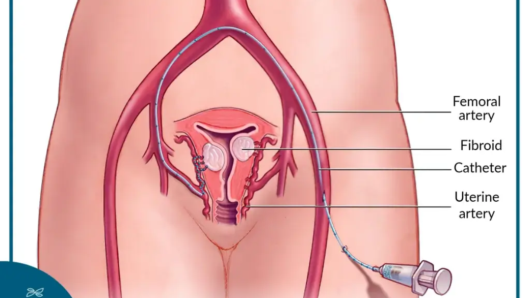
A primary interventional radiology technique used for treating uterine fibroids is Uterine Fibroid Embolization (UFE). During this procedure, an interventional radiologist makes a tiny incision in the skin, and they insert a catheter into the artery. Using real-time imaging, the doctor guides the catheter to the uterine arteries that supply blood to the fibroids.
Once in place, small particles are injected into the uterine arteries to block the blood flow to the fibroids. Deprived of blood, the fibroids shrink and die over time, and this leads to a reduction in symptoms like heavy bleeding and pain. Consulting a specialist helps determine the right technique for each situation.
Treat Uterine Fibroids Now
Addressing uterine fibroids promptly can improve quality of life and prevent complications such as severe anemia. With advances in interventional radiology, patients have access to effective, minimally invasive treatments that enable quick recovery and preserve the uterus. Consult a specialist and learn more about your treatment options today.